DNA Compaction Unraveled: Cellular Factories Revealed
The human body is a marvel of microscopic engineering. One of its most impressive feats is how it manages the vast amount of genetic information within each cell. Consider this: every cell contains approximately two meters of DNA, which must be meticulously packed into a nucleus that is merely a fraction of the width of a human hair. This incredible compression must be achieved without tangling the DNA or rendering it inaccessible for vital cellular processes like reading genes.
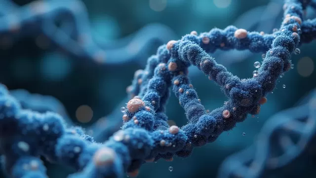
The initial stages of this process are well-understood. The DNA strand is first wound around protein spools, creating structures called nucleosomes. These nucleosomes are then strung together and folded into more compact chromatin fibers. For years, however, the final step in this extreme condensation remained a puzzle for scientists.
Unveiling a New State of Matter in the Cell
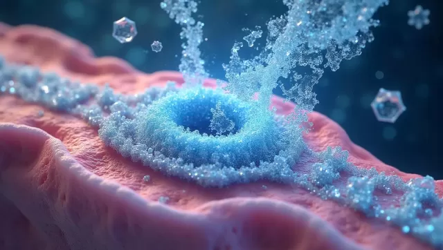
A major breakthrough came when researchers, led by Michael Rosen at UT Southwestern Medical Center, discovered that lab-created nucleosomes could spontaneously self-organize into dense, liquid-like droplets. This process, known as phase separation, is chemically similar to how oil forms distinct beads in water. These structures, called biomolecular condensates, are formed without a surrounding membrane and are believed to mirror how chromatin achieves its ultimate dense state within a living cell.
These condensates are dynamic environments, composed of hundreds of thousands of individual molecules in constant motion. Their collective behavior gives rise to unique physical properties, or emergent properties, that are not present in the isolated molecules. These group dynamics dictate how the droplets are formed, their consistency, and their function. To truly grasp how this works, scientists needed a way to peer deep inside these droplets and see the molecular arrangement for themselves.
High-Resolution Imaging Solves a Biological Puzzle
A collaborative effort involving Rosen's team, Elizabeth Villa at the University of California, San Diego, and other leading researchers has now accomplished this. Leveraging state-of-the-art imaging technology at the Janelia Research Campus, the team captured unprecedentedly detailed images of the internal architecture of these synthetic chromatin condensates.
By integrating these high-resolution snapshots with advanced computer simulations, the researchers were able to map out the molecular interactions and structures within the droplets. Their findings revealed a critical detail: the length of the flexible "linker DNA" that connects one nucleosome to the next plays a decisive role in the overall organization. This arrangement directly influences how the chromatin fibers interact with each other, shaping the internal network of the entire condensate.
This discovery explains why certain types of chromatin can undergo phase separation more readily than others and why condensates made from different chromatin building blocks possess distinct material characteristics. Crucially, the team also confirmed that the structure of these lab-made condensates bore a striking resemblance to the highly compacted chromatin observed inside actual cells. This work successfully connects the behavior of single molecules to the macroscopic properties of the larger assemblies they form.
Broader Implications for Health and Disease
The insights gained from this research extend far beyond DNA packaging. The methodologies developed provide a powerful new framework for studying the vast array of biomolecular condensates found in cells. These membrane-less organelles are critical for a wide range of cellular activities, from controlling gene expression to managing cellular stress.
Understanding the principles of condensate assembly is also vital for medicine. A growing body of evidence suggests that disruptions in this process, or abnormal condensation, may be a contributing factor in a host of human diseases, including neurodegenerative conditions like ALS and certain types of cancer. By deciphering how these structures form and function, researchers hope to understand what goes wrong in disease states. As lead author Huabin Zhou explains, this fundamental knowledge could pave the way for an entirely new class of therapeutics designed to correct or target these cellular processes.