Lab-Grown Embryo Models Yield Human Blood Cells
Groundbreaking research has achieved a significant milestone in regenerative medicine: the successful cultivation of embryo-like structures in a laboratory setting that are capable of producing human blood cells. This breakthrough paves the way for potential treatments involving patient-specific bone marrow transplants and offers unprecedented insights into the earliest stages of human development.
The research team, led by Dr. Jitesh Neupane at the University of Cambridge's Gurdon Institute, meticulously crafted these embryo models using human stem cells, circumventing the need for eggs or sperm. The self-organizing structures mimic the natural developmental processes, allowing scientists to observe the formation of various cell types.
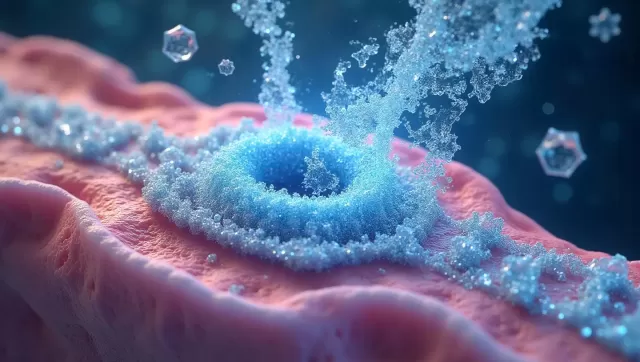
"Seeing the emergence of the blood-red color was an exhilarating moment," Dr. Neupane noted, emphasizing the visual confirmation of their success.
The applications of this research are vast, ranging from drug screening and the study of early blood and immune system development to modeling blood disorders like leukemia. The ability to generate blood stem cells that are fully compatible with a patient's own body has transformative potential for personalized medicine.
Existing methods for creating human blood stem cells often rely on complex cocktails of proteins. This novel approach stands out by replicating the natural embryonic developmental process, creating a more authentic and efficient pathway.
Professor Azim Surani, the senior author of the paper, emphasized the significance of this advancement: "The capacity to produce human blood cells in the lab represents a major stride towards regenerative therapies, harnessing a patient's own cells to repair and regenerate damaged tissues."
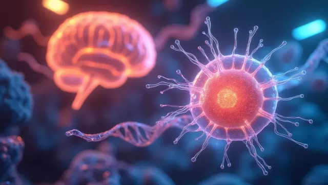
The embryo models were designed with a crucial distinction: they lack the tissues that develop into the placenta and yolk sac in a natural embryo. This intentional design prevents the potential for the model to develop into a fetus and excludes the formation of brain tissue, addressing ethical considerations.
"This is a highly refined system," explained Dr. Neupane.
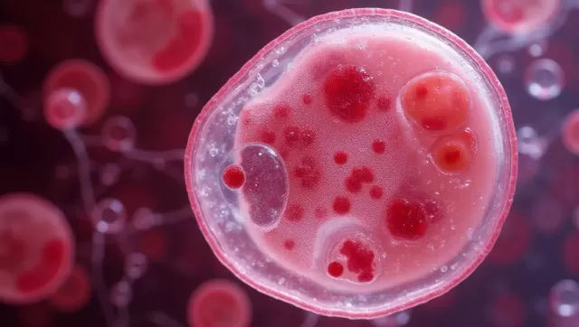
Under microscopic observation, the team witnessed the remarkable self-organization of the stem cells into three germ layers – ectoderm, mesoderm, and endoderm – within just two days. These layers form the foundational structure of the human body. By day eight, the model exhibited beating heart cells, mirroring the development of the heart in a natural embryo.
Around day 13, distinct red patches of blood appeared, indicating the presence of blood cells. Further analysis confirmed that the blood stem cells derived from the model could differentiate into various blood cell types, including oxygen-carrying red blood cells and crucial immune system white blood cells.
These remarkable findings, published in Cell Reports, mark a pivotal step forward in our understanding of early human development and offer immense promise for future regenerative medicine therapies.